Herbivores
Be sure to write about what you are learning in the lab section of your notebook. You will be expected to answer questions about the lab activity during the lab self test and lab quiz. It helps to have your text and coloring books open beside you for support.
| Refer to the Assigned Readings Below: | |
| Marine Biology Textbook | Chapter 10, page 219 to 220 |
| Marine Biology Coloring Book | Plate 29, 30, 31, 35, 106 and 107 |
Predators
Predators are animals which capture, kill and consume other organisms which are called prey. The prey organism can be of any type. Predators are classified by the type of prey they cconsume.
Herbivores
Predators that eat plants, seaweeds and algae are called herbivores. There are two types of herbivores: macrophagous and microphagous. Macrophagous herbivores eat large plants and seaweeds or parts of large plants and seaweeds. Microphagous herbivores eat small algae and seaweeds which they must gather in large numbers. Microphagous herbivores collect their food in different ways. Some are grazers that scrape algae from surfaces and some are filter-feeders that filter it out of the water.
|
Top Snails
|
|
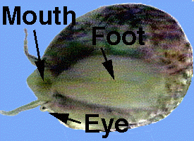
| Top snails are microphagous herbivores. They are grazers that collect their food by scraping it from rocks with a special chitinous "tongue" called a radula. The radula is a file-like structure that has many rows of teeth. The radula is extended through the mouth and applied to the rock and then retracted back into the mouth. On the return trip the teeth of the radula scrape algae into the mouth. | |
Experimental
Set Up:
|
|
|
Observations:
|
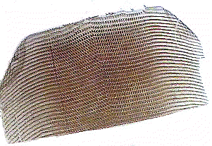
Examine the photograph of the Top Snail and note the radula in it's mouth. |
|
Snail Radula
|
||
| The teeth on the radula of a snail are specific for the type of food it eats. In fact the radulae of different snail species are structurely so different that they can be used to identify the snails. | ||
Experimental
Set Up:
|
||
|
Observations:
|
Examine the photograph of the snail radula and note the arrangement of its teeth. |
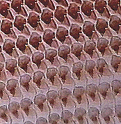
Examine the photograph of the snail radula and note the arrangement of its teeth. |
|
Acorn Barnacles
|
|
|
Acorn Barnacles are microphagous herbivores that collect their food by filtering it out of the water using their cirri (legs). The cirri are held together in the shape of a basket as they are swept through the water. Algal cells are captured on the cirri and are transferred to the mouth at the base of the basket. |
|
Experimental
Set Up:
|
|
|
Observations:
|
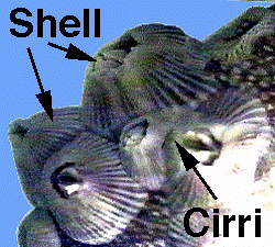
Examine the photograph of the Acorn Barnacles and note the cirri extending from one's shell. |
|
Stalked Barnacles
|
|
|
Stalked barnacles are stalked barnacles that feed in the same way as acorn barnacles. They are easier to examine and photograph than acorn barnacles because of their larger size and the arrangement of their shell plates and soft parts. |
|
Experimental
Set Up:
|
|
|
Observations:
|

Examine the photograph of the stalked barnacle and carefully observe the cirri. |
|
Mussels
|
|
|
Mussels are microphagous herbivores that collect their food by filtering it out of the water using their gills. Water is pumped into the cavity inside the shell by the beating of many tiny, hair-like cilia on the gills and the thin, fleshy mantle that lines the shell. The water passes through the gills and exits from the shell cavity. Algae is filtered from the water by the gill and is conducted by cilia along the gill toward the mouth. At the top of the gill a set of small labial palps pass the food from the gill to the mouth. |
|
Experimental
Set Up:
|
|
|
Observations:
|
Carefully observe the action of the gills as it moves the carmine particles. |
|
Position of Mussel Gills
|
|
|
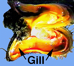
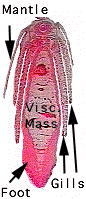
The paired gills of the mussel are arranged on either side of its visceral mass. They hang down into the shell cavity. |
|
Experimental
Set Up:
|
|
|
Observations:
|
Examine the photograph of the mussel cross section and note the position of its gills to the right and left of the central visceral mass and foot. |
|
Ciliary Action of Mussel Gills
|
|
|
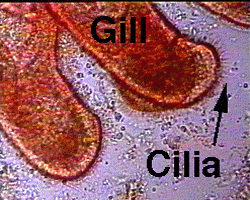
The cells that cover the filaments of the mussel's gill have microscopic hairs called cilia which beat to move particles on the gill surface. |
|
Experimental
Set Up:
|
|
|
Observations:
|
Carefully observe the cilia. |
Lab Activity 7.4 Carnivores and Saprophages