Shark Urogenital and Nervous Anatomy
Be sure to write about what you are learning in the lab section of your notebook. You will be expected to answer questions about the lab activity during the lab self test and lab quiz. It helps to have your text and coloring books open beside you for support.
| Refer to the Assigned Readings Below: | |
| Marine Biology Textbook | Chapter 8, pages 166 to 169 and 174 to 175 |
| Marine Biology Coloring Book | Plates 49 to 51 |
|
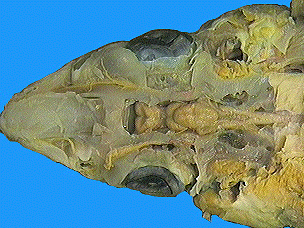
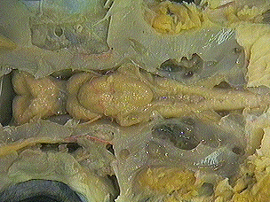
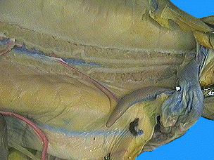
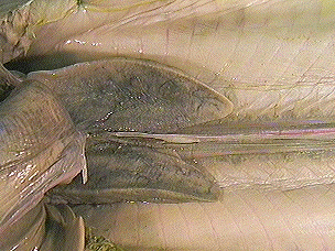
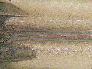
The specimen in the phographs was prepared by removing almost the entire liver, alimentary canal, pancreas, and spleen. This revealed the urogenital structures: gonads, kidneys, and associated ducts. The urinary and genital systems have distinct and unique functions: (1) the removal of nitrogenous wastes and the maintenance of water balance, and (2) the reproduction of the species. However, due to their similar developmental origins and common structures, they are usually considered as a single system. The shark kidney and its ducts are quite different from those in higher vertebrates. The relationship between the urinary and genital structures is also quite different. The kidneys are flattened, ribbon-like, darkly colored structures lying dorsally on either side of the midline, along the entire length of the body cavity. A tough, white, glistening strip of connective tissue is found between the kidneys in the midline. The kidneys of the male are essentially the same as those of the female. The posterior portion is involved in the manufacture and transport of urine. The main difference lies in the anterior portion of the kidney, which in females has no function, but in males is an active part of the reproductive system.
|
|
|
In males, paired testes lie near the anterior end of the body cavity, dorsal to the liver, adjacent to the anterior ends of the kidneys. The sperm pass from the testes to the kidneys within narrow tubules called efferent ductules. |
|
|
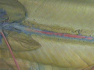
After passing through the anterior end of the kidney (in males) the sperm enter the ductus deferens and pass posteriorly toward the cloaca. In mature male specimens, the ductus deferens may be seen on the ventral surface of the kidneys as a pair of highly coiled tubules. In females this duct carries urine. In males it transports spermatozoa and seminal fluid. In males, the posterior portion of the ductus deferens widens and straightens to form the paired seminal vesicles. |
|
|
In males, the paired sperm sacs at the posterior ends of the seminal vesicles receive the seminal secretions. They join to form the urogenital sinuses, which exit through the fleshy conical urogenital papilla which extends from the cloaca. The accessory urinary ducts collect and transport urine from the kidneys. These paired thin tubules may be found along the medial side of the posterior half of the kidney. Small collecting tubules from the kidneys lead into the accessory urinary ducts along their lengths. The cloaca receives the genital and urinary products as well as the feces. |
|
|
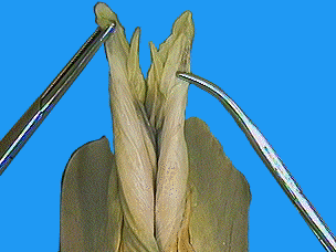
The claspers are modified extensions of the medial portions of the pelvic fins. They are inserted into the female's cloaca during copulation. The finger-like claspers each have a dorsal groove, the clasper tube that carries the seminal fluid from the cloaca of the male to the cloaca of the female during mating. |
|
|
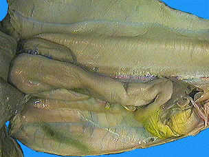
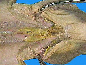
The specimen in the phographs was prepared by removing almost the entire liver, alimentary canal, pancreas, and spleen. This revealed the urogenital structures: gonads, kidneys, and associated ducts. In females, the ovaries are two cream-colored elongated organs in the anterior part of the body cavity, dorsal to the liver on either side of the mid-dorsal line. The shape of the ovaries will vary depending upon the maturity of the specimen. In immature females, they will be undifferentiated and glandular in appearance. In mature specimens, you may find two to three large eggs, about three centimeters in diameter, in each ovary. When these break the surface of the ovary, upon ovulation, they enter the body cavity and by means of peritoneal cilia are moved into the oviducts. |
|
| The
oviducts are elongated tube-like structures lying dorsolaterally
the length of the body cavity, along the sides of the kidneys. In
mature specimens they are more prominent. The distal half of the oviduct
is enlarged to form the uterus.
The shell gland is the anterior end of the oviduct. The eggs are fertilized and receive a light shell-like covering as they pass through the shell gland. |
|
|
The posterior half of the oviduct becomes enlarged and is known as the uterus. The fertilized eggs develop into embryos in the uterus. Upon completing their period of gestation (close to two years), the young are ready to be born. The cloaca serves for the elimination of urinary and fecal wastes as well as an aperture through which the young "pups" are born. The two uteri open into the posterodorsal portion of the cloaca just ventral to the urinary papilla. Fertilization in the female dogfish shark is internal, usually taking place within the shell gland of the oviduct. The fertilized eggs continue to move posteriorly to the uterus. As they grow the pups are attached to the egg, now known as the yolk sac, by means of a stalk. During its period of gestation, which is nearly two years, the yolk is slowly absorbed by the shark "pup." Numerous uterine villi, finger-like projections from the uterine wall, make contact with the surface ot the developing embryo and its yolk sac. It is believed that these provide the embryo with water; all other nutrients are supplied by the yolk. At birth the young are about 23 to 29 centimeters long. This type of development, where the young are born as miniature adults but have received hardly any nutrition directly from the mother's uterus, is known as ovoviviparous. |
|
|
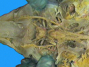
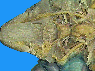
The specimen in the photographs was prepared by removing the skin from the dorsal surface of the head and shaving off thin horizontal chips of cartilagenous cranium until the brain and cranial nerves were exposed. The nervous system functions in communication between the various parts of an organism and between the organism and its external environment. It consists of the central nervous system (brain and spinal cord) and the peripheral nervous system (sense organs, cranial and spinal nerves). |
|
|
Forebrain
Midbrain
Hindbrain
|
|
|
Cranial Nerves The cranial nerves originate in the brain and exit at the chondrocranium. These nerves can be sensory, carrying impulses to the brain; they can be motor, carrying impulses from the brain to muscles and glands; or they can be mixed nerves, carrying both sensory and motor fibers. The cranial nerves of all vertebrates have similar names and similar functions. Fish are usually described as having ten pairs of cranial nerves, including:
|
|
|
The olfactory sacs are spherical structures that contain a series of radial folds called olfactory lamellae. Their surfaces are covered with olfactory epithelium. Sea water taken into the nares is passed over these sensory areas. Here the odors stimulate the cilia-like endings of neuro-sensory cells. The olfactory bulbs are a paired anterior extension of the brain leading into the posterior end of the olfactory sacs. Their fibers continue into the olfactory tract and the olfactory lobe of the cerebral hemisphere. The sclera is the tough, white, fibrous outer coat of the eye. At places it is made even more firm by cartilage embedded in the sclera. The iris is the pigmented anterior extension of the choroid layer. In its center is the pupil. The iris regulates the size of the pupil. In the living shark the lens is a clear and flexible structure located behind the iris. It helps to focus the light upon the light sensitive retina. The retina is the multi-layered sensory gray-white colored membrane. The rods and cones which receive light stimuli are located here. The optic nerve leaving the eye is a continuation of the light receptor cells in this membrane. |
|