Shark Digestive Anatomy
Be sure to write about what you are learning in the lab section of your notebook. You will be expected to answer questions about the lab activity during the lab self test and lab quiz. It helps to have your text and coloring books open beside you for support.
| Refer to the Assigned Readings Below: | |
| Marine Biology Textbook | Chapter 8, page 164 |
| Marine Biology Coloring Book | Plates 49 to 51 |
|
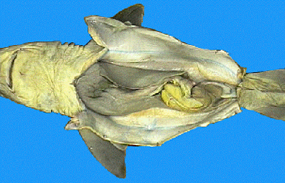
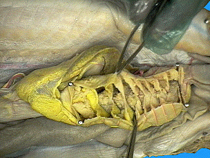
The shark specimen in the photographs was prepared by turning it ventral side up and making a mid-ventral incision just anterior to the cloacal opening. It was cut in an anterior direction slightly to the right of the mid-ventral line. This cut was continued all the way to the pectoral girdle. The inside of the large body cavity was exposed. The large flaps of body wall were folded back and pinned. A smooth, shiny membrane called peritoneum can be seen lining the inside of the body wall. The visceral organs are suspended dorsally by a double membrane of peritoneum know as mesentery. The liver is the largest organ lying within the body cavity. Its two main lobes, the right and left lobes, extend toward the posterior from the pectoral girdle most of the length of the cavity. A third, much shorter lobe is located medially and contains the green gall bladder along its right edge. The liver is rich in oil which stores energy for the shark. The oil's low specific gravity is also responsible for giving the shark a limited amount of buoyancy.
|
|
|
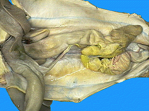
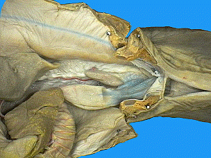
The shark specimen in the photographs was prepared by moving the large liver to the shark's right side. The esophagus is the thick muscular tube extending from the top of the cavity connecting the oral cavity and pharynx with the stomach. The esophagus leads into the J-shaped stomach. The upper portion, the cardiac region, continues as the main body and ends at the duodenal end. |
|
|
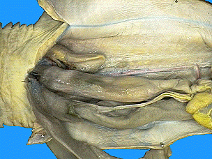
The shark specimen in the photographs was prepared by cutting the stomach open along its long axis. This shark's stomach contained the partially digested remains of fish, which were washed out under running water. The mucosa is the inner lining of the stomach. The rugae are longitudinal folds that help in the churning and mixing the food with digestive juices. A circular muscular valve, the pyloric sphincter, is located at the far end or pyloric end of the stomach. It regulates the passage of partially digested food into the intestines. |
|
|
The specimen in the photographs was prepared by moving the large liver forward. The duodenum is a short U-shaped portion of the small intestine that connects the stomach to the intestine. The bile duct from the gall bladder enters the duodenum. The pancreas is located on the duodenum and the lower stomach. The secretions of the pancreas enter the duodenum by way of the pancreatic duct. The dark, triangular-shaped spleen is located near the posterior end of the stomach. Although a part the lymphatic system, the spleen is closely associated with the digestive organs in all vertebrates. The valvular intestine is the second and much larger portion of the small intestine. It follows the duodenum and its outer surface is marked by rings. |
|
|
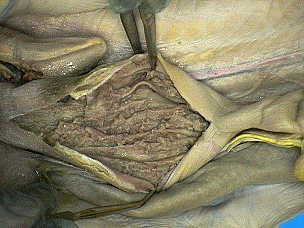
The shark specimen in the photographs was prepared by cutting away the outer tissue of the valvular intestine. The spiral valve is the screw-like, symmetrical shape within the valvular intestine. It adds surface area for digestion and absorption to an otherwise relatively short intestine. |
|
|
The shark specimen in the photographs was prepared by pulling the intestine forward. The colon is the narrowed continuation of the valvular intestine. It is located at the posterior end of the body cavity. The rectal gland is a slender, blind-ended, finger-like structure that leads into the colon by means of a duct. It has been shown to excrete salt (NaCI) in concentrations higher than that of the shark's body fluids or sea water. It is thus an organ of osmoregulation, regulating the shark's salt balance. The cloaca is the last portion of the alimentary canal. It collects the products of the colon as well as the urogenital ducts. It is a catch-all basin leading to the outside by means of the cloacal opening. |
|
Lab Activity 5.3 Shark Respiratory and Circulatory Anatomy